 Products Home
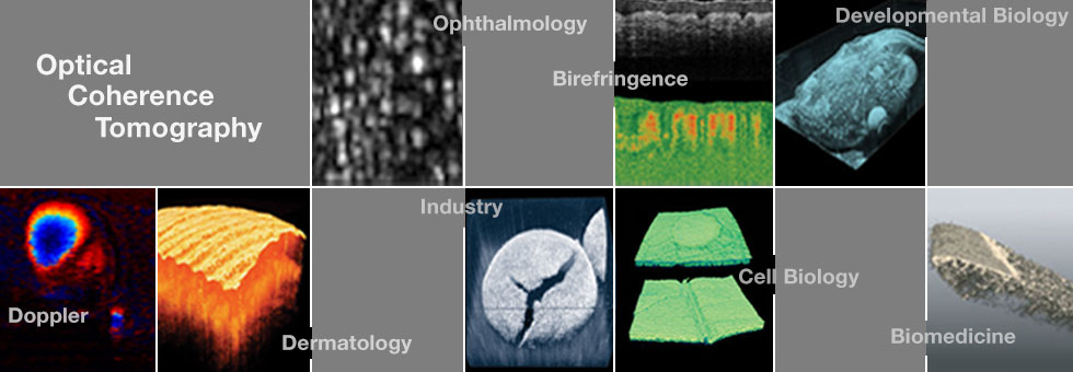

| Developmental Biology |

Live imaging of blood flow in mammalian embryos using Doppler swept-source optical coherence tomography
I. V. Larina, et al., Journal of Biomedical Optics, 13 (6), 2008.
Link

Doppler optical cardiogram gated 2D color flow imaging at 1000 fps and 4D in vivo visualization of embryonic heart at 45 fps on a swept source OCT system
A. Mariampillai, et al., Optics Express, 15 (4), 2007.
Link
| Vascular Imaging |

Rapid volumetric angiography of cortical microvasculature with optical coherence tomography
V. J. Srinivasan, et al., Optics Letters, 35 (1), 2010.
Link

Speckle variance detection of microvasculature using swept-source optical coherence tomography
A. Mariampillai, et al., Optics Letters, 33 (13), 2008.
Link

Blood-vessel closure using photosensitizers engineered for two-photon excitation
H. A. Collins, et al., Nature Photonics, 2008.
Link
| Biomedical |

Swept source optical coherence microscopy using a 1310 nm VCSEL light source
O. O. Ahsen, et al., Optics Express, 21 (15), 2013.
Link

MEMS tunable VCSEL light source for ultrahigh speed 60kHz - 1MHz axial scan rate and long range centimeter class OCT imaging
B. Potsaid, et al., Proc. SPIE 8213, doi:10.1117/12.911098, 2012.
Link

Ultrahigh speed endoscopic optical coherence tomography using micro-motor imaging catheter and VCSEL technology
T. H. Tsai, et al., Proc. SPIE 8571, doi:10.1117/12.2006952, 2013.
Link

High-speed ultra-broad tuning MEMS-VCSELs for imaging and spectroscopy
V. Jayaraman, et al., Proc. SPIE 8763, doi:10.1117/12.2018345, 2013.
Link

Ultrahigh speed endoscopic optical coherence tomography using micromotor imaging catheter and VCSEL technology
T. H. Tsai, et al., Biomedical Optics Express, 4 (7), 2013.
Link

Phase-sensitive swept-source optical coherence tomography imaging of the human retina with a vertical cavity surface-emitting laser light source
W. Choi, et al., Optics Letters, 38 (3), 2013.
Link

Automated quantification of microstructural dimensions of the human kidney using optical coherence tomography (OCT)
Q. Li, et al., Optics Express, 17 (18), 16000, 2009.
Link

Optical coherence tomography (OCT) reveals depth-resolved dynamics during functional brain activation
Y. Chen, et al., Journal of Neuroscience Methods, 178 (1), 2009.
Link

Three-dimensional coregistered optical coherence tomography and line-scanning fluorescence laminar optical tomography
S. Yuan, et al., Optics Letters, 34 (11), 2009.
Link

High-resolution optical coherence tomography of the living kidney
P. M. Andrews, et al., Laboratory Investigation, 88, 2008.
Link

Optical coherence tomography as an orientation guide in cochlear implant surgery?
H. W. Pau, et al., G. Acta Oto-Laryngologica, 127 (9), 2007.
Link

Combined impedance spectroscopy and Fourier domain optical coherence tomography to monitor cells in three-dimensional structures
P. O. Bagnaninchi, International Journal of Artificial Organs, 33 (4), 2010.
Link

Enhanced optical clearing of skin in vivo and optical coherence tomography in-depth imaging
X. Wen, et al., Journal of Biomedical Optics, 17 (6), 2012.
Link
| Industrial |

Application of optical coherence tomography to automated contact lens metrology
B. R. Davidson, et al., Journal of Biomedical Optics, 15 (1), 2010.
Link

Imaging Pharmaceutical Tablets with Optical Coherence Tomography
J. M. A. Mauritz, et al., Journal of Pharmaceutical Sciences, 99 (1), 2009.
Link

Comparison of three-dimensional optical coherence tomography and high resolution photography for art conservation studies
D. C. Adler, et al., Optics Express, 15 (24), 2007.
Link
| Ophthalmology |

Retinal, anterior segment and full eye imaging using ultrahigh speed swept source OCT with vertical- cavity surface emitting lasers
I. Grulkowski, et al., Biomedical Optics Express, 3 (11), 2012.
Link

Ophthalmic Applications of Ultrahigh Speed OCT Using VCSEL Light Source Technology
I. Grulkowski, et al., Invest. Ophthalmol. Vis. Sci., 53, 2012.
Link

Reproducibility of a Long-Range Swept-Source Optical Coherence Tomography Ocular Biometry System and Comparison with Clinical Biometers
I. Grulkowski, et al., Ophthalmology, doi:10.1016/j.ophtha.2013.04.007, 2013.
Link

Handheld High Speed 500 kHz Swept Source OCT Device Using a Micro Scanning Mirror
C. Lu, et al., Invest. Ophthalmol. Vis. Sci., 54, 2013.
Link

VCSEL Laser Technology for Ultrahigh Speed and Extended Depth Range OCT Imaging of the Retina and Anterior Eye
B. Potsaid, et al., Invest. Ophthalmol. Vis. Sci., 54, 2013.
Link
Three-Dimensional Biometric Measurements of Accommodation Using Full-Eye-Length Swept-Source OCT
I. Grulkowski, et al., Invest. Ophthalmol. Vis. Sci., 54, 2013.
Link

Ultrahigh speed polarization sensitive OCT of the anterior and posterior eye using a 1050 nm VCSEL light source
A. H. Dhalla, et al., Invest. Ophthalmol. Vis. Sci., 54, 2013.
Link

Handheld High Speed Swept Source Optical Coherence Tomography at 1050nm
C. D. Lu, et al., Invest. Ophthalmol. Vis. Sci., 53, 2012.
Link

Ultrahigh speed Spectral / Fourier domain OCT ophthalmic imaging at 70,000 to 312,000 axial scans per second
B. Potsaid, et al., Optics Express, 16 (19), 2008.
Link

High-speed, high-resolution optical coherence tomography retinal imaging with a frequency-swept laser at 850 nm
V.J. Srinivasan, et al., Optics Letters, 32 (4), 2007.
Link

Imaging thermal expansion and retinal tissue changes during photocoagulation by high speed OCT
H. H. Müller, et al., Biomedical Optics Express, 3 (5), 2012.
Link
| Other Publications |

Space-division multiplexing optical coherence tomography
C. Zhou, et al., Optics Express, 21 (16), 2013.
Link

High-precision, high-accuracy ultralong-range swept-source optical coherence tomography using vertical cavity surface emitting laser light source
I. Grulkowski, et al., Optics Letters, 38 (5), 2013.
Link

Binary-phase spatial filter for real-time swept-source optical coherence microscopy
L. Liu, et al., Optics Letters, 32 (16), 2007.
Link

Three-dimensional and C-mode OCT imaging with a compact, frequency swept laser source at 1300 nm
R. Huber, et al., Optics Express, 13 (26), 2005.
Link
| Posted Comments: | |
| No Comments Posted |